Age Related Macular Degeneration (ARMD)
The retina is a multi-layered structure lining the inside of the back of the eye that picks up the incoming light as an image and sends it to the brain. Age-related macular degeneration (ARMD) is the retina wearing out. It takes two forms – dry ARMD and wet ARMD. Dry ARMD is a progressive degeneration of the retina, slowly reducing vision. Wet ARMD is an abnormal blood vessel developing under the retina that leaks or bleeds and results in a rapid reduction and distortion of vision.

Astigmatism
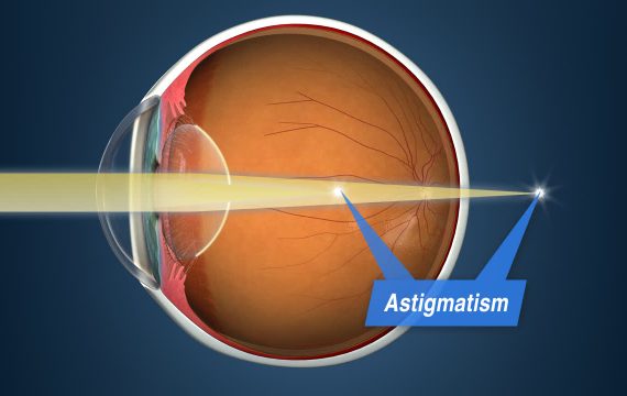
Astigmatism describes an irregular corneal surface. It is steeper in one direction compared to another, like the back of a spoon. Like short or long sight, astigmatism describes the refractive error. It is not an eye disease or eye health problem, just an issue with how the eye focuses light. In an eye with astigmatism, there are multiple focus points (either in front of the retina, behind it, or both). It cannot produce a clear image so patients with experience blurred vision at all distances. Patients with astigmatism can be free from glasses and contact lenses with a procedure that suits their age:
- For those aged 20-45 years, LASIK is preferred, while an intracollamer lens (ICL) may suit those with a thin cornea or high prescription.
- For those people aged over 45 years, refractive lens exchange with premium intraocular lens implants corrects vision, removes the lens so a cataract will never develop and restores youthful vision for life.
- While those who are 60+ years of age, cataract surgery with premium intraocular lens implants corrects vision, removes the cataract and restores youthful vision for life.
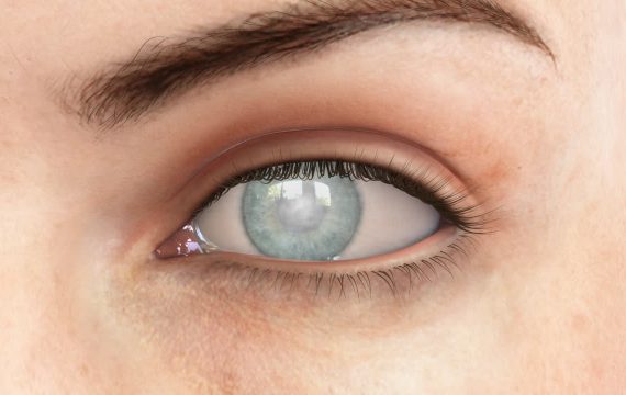
Cataract
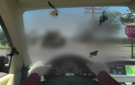
A cataract is clouding of the lens in one or both eyes. As the natural lens ages, it goes from crystal clear and flexible to hard and less transparent. Cataracts are part of the normal aging process and one of the most common causes of vision loss as we age. With cataracts, vision becomes blurry and images are not as bright or colourful as they used to be, the eyes can become more sensitive to glare from the sun or oncoming headlights. Daily activities such as reading, driving and computer work are difficult without the aid of glasses. Cataract surgery is a simple, painless procedure to regain vision and is the only effective treatment currently available. There are no medications or eye drops that can halt or reverse this process. In cataract surgery, the clouding lens is replaced with a clear artificial implant. Freedom Eye Laser utilises the latest generation of lens implants that correct for your prescription to eliminate the need for glasses at all distances for life. Find out more...

Dermatochalasis
Dermatochalasis is a medical condition that develops as part of the normal aging process and typically occurs in patients over 50 years of age. Otherwise known as “baggy eyes”, dermatochalasis describes an excess skin of the upper and lower eyelids. It occurs as the underlying connective tissue supporting the skin weakens, allowing the skin to stretch and sag. In dermatochalasis, the upper lid hoods over the eye, obscuring vision and creating a sensation of heaviness. The person elevates their forehead to clear the visual axis, resulting in brow ache, fatigue and headaches. Cosmetically, dermatochalasis increases the appearance of age and tiredness. It is corrected with a blepharoplasty, a surgical procedure that removes this skin for either functional, visual or cosmetic reasons.
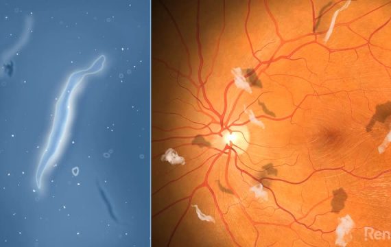
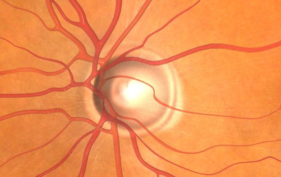
Diabetic Retinopathy
Elevated blood sugar levels in the context of diabetes can cause progressive damage to the retinal blood vessels. This damage causes the blood vessels to either leak or block off. If the vessel leaks, the released fluid causes the retina to swell and vision is reduced. If the vessel blocks off, the reduced blood supply to the retina causes the growth of new, abnormal blood vessels that can bleed. The blood released into the vitreous (the jelly filling the back of the eye) profoundly reduces vision. Subsequent fibrosis can then cause the retina to detach resulting in blindness. As the early changes from diabetes in the eye have no symptoms, every person with a diagnosis of diabetes should be monitored as early treatment can prevent ongoing damage.
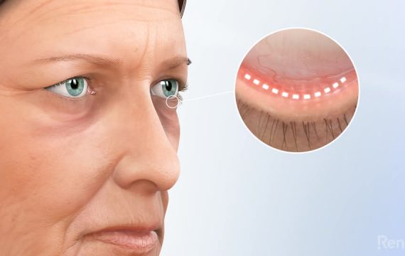
Dry Eye Syndrome
Dry eye syndrome is an extremely common condition that can cause irritation and can blur vision. It is now possible to perform advanced diagnostic tests to isolate the cause of an individual’s dry eye and target therapy to improve treatment effectiveness. At Freedom Eye Laser we have invested in advanced diagnostics and treatment options to provide a dry eye centre of excellence. Our speciality clinic supports people experiencing this chronic, debilitating condition. The most common cause of dry eye is dysfunction of the meibomian glands in the lid margin. These glands produce the oily layer of the tears. If they get inflamed, they do not work properly and you get a reduction in this oily layer. This, in turn, allows for accelerated tear evaporation and subsequent dry eye. Certain environments such as wind or air conditioning can exacerbate these symptoms. Paradoxically it can cause a watery eye that’s dry in response to the unstable tear film. Other causes of dry eye include Blepharitis (is an inflammatory condition of the eyelid margin that is a common cause of ocular discomfort and irritation at all ages); an autoimmune reduction in tear production (Sjogren’s Syndrome); and allergic eye disease to environmental irritants such as dust, pollens or animal fur. Discover how Freedom Eye Laser offer tailored solutions to achieve relief from dry eye syndrome. Learn more...
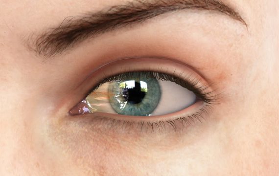
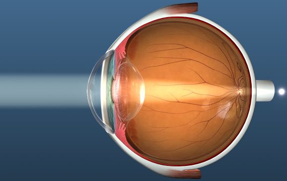
Emmetropia
Emmetropia means normal sightedness, or perfect vision. The cornea and the crystalline lens within the eye, work together to focus light on the retina to register a sharp image which is projected to the brain. Emmetropia is the goal of any refractive surgery, resulting in crisp clear vision. Freedom Eye Laser is expert at achieving this.
Floaters
The vitreous (the gel that fills the back of the eye) becomes more of a liquid as you age. The reduced volume of the liquid pockets results in the vitreous eventually collapsing into itself. Flashes can be experienced at this point due to traction on the retina stimulating the photoreceptors. The vitreous pulls off from its attachment to the optic disc, taking a fragment of tissue with it that then floats in front of the retina. Light hits the tissue fragment and it throws a shadow onto the retina, experienced as a black floater. Occasionally the pull of the vitreous is strong enough to tear the retina. If the tear is not treated, fluid can enter through the tear and under the retina, detaching it. If a retinal detachment is untreated blindness will result. Although people often get used to floaters, they can be very symptomatic. Vision can be constantly interrupted which is distracting and frustrating.
Fuchs Endothelial Dystrophy
Fuchs Endothelial Dystrophy is an inherited condition that causes progressive visual blurring. The cornea is the clear dome of tissue on the front of the eye. Light enters through the cornea, which focuses the light rays so that you can see clearly. The cornea needs to be relatively dehydrated to maintain transparency. The inner lining of the cornea is a single layer of cells called the endothelium. Its function is to pump fluid from the cornea back inside the eye to maintain this dehydration. Endothelial cells do not replicate. Over time, every person progressively loses these cells. A normal cornea has enough cells to last a lifetime, however people with Fuchs Endothelial Dystrophy are born with a reduced number of cells and eventually reach a point where they cannot dehydrate their cornea. The cornea becomes waterlogged and misty, requiring treatment.
Glaucoma
The front of your eye is filled with fluid called the aqueous humour that provides nutrients to the internal structure. The pressure this fluid creates gives the eye its structural stability, similar o the air in a balloon. This fluid is drained through a meshwork of tissue; if this meshwork is too tight there is an increase in pressure to force the fluid out. This leads to an elevation of the internal eye pressure. The pressure elevation progressively compresses and damages the retina causing blind spots. Without treatment this eventually results in blindness. Over the age of 50 years, everyone should have their eye pressures checked, particularly if there is a family history of glaucoma. In the early stages there are no symptoms detectable by the patient despite the damage occurring so regular check ups are therefore critical.
Hyperopia
Without glasses or contacts, patients with hyperopia experience blurred vision up close, while objects in the distance are usually clearer. In some cases, both distance and close vision can lack focus. Long-sightedness, just like short-sightedness and astigmatism describe refractive error. This means that it is not an eye disease or eye health problem, but an issue with how the eye focuses light. Hyperopia occurs when the cornea and lens curvature is too flat for the length of the eye so that the image focuses at a point behind the retina. Patients with hyperopia can be free from glasses and contact lenses with surgery that typically suits their age. For those aged 20-45 years, LASIK is preferred while an intracollamer lens (ICL) may better suit those with a thin cornea or high prescription. For people 45 years+ refractive lens exchange with premium intraocular lens implants corrects vision, removes the lens so a cataract will never develop and restores youthful vision for life. For people who are 60+ years of age, cataract surgery with premium intraocular lens implants corrects vision, removes the cataract and restores youthful vision for life.
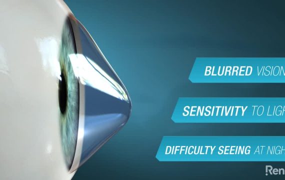
Keratoconus
Keratoconus literally means “cone-shaped cornea”. The cornea is the clear dome of tissue on the front of the eye. Light enters through the cornea, which focuses the light rays so that you can see clearly. Keratoconus is an inherited condition where the clear front surface of the eye, the cornea, is excessively elastic and with time starts to distort. The cornea ends up looking like the side of a pear, resulting in an asymmetry that causes visual distortion and blurred vision. It naturally progresses with time unless it can be stopped with treatment.
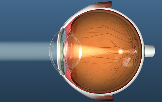
Myopia
Without glasses or contacts, patients with myopia experience blurred vision in the distance, while closer objects are clear and focused. Short-sightedness, like farsightedness and astigmatism, describes refractive error. This means that it is not an eye disease or eye health problem, just an issue with how the eye focuses light. Myopia occurs when the cornea and lens curvature are too steep for the length of the eye so the image focuses at a point in front of the retina, leading to blurred distance vision. Patients with myopia can be free from glasses and contact lenses with surgery that typically suits their age. For those aged 20-45 years, LASIK is preferred, while an intracollamer lens (ICL) may suit those with a thin cornea or high prescription. For those people aged over 45 years, refractive lens exchange with premium intraocular lens implants corrects vision, removes the lens so a cataract will never develop and restores youthful vision for life. While those who are 60+ years of age, cataract surgery with premium intraocular lens implants corrects vision, removes the cataract and restores youthful vision for life.
Pinguecula
A pinguecula (pronounced pin- GWEK-yoo-la) is a nodule or elevation on the white of the eye that is caused by UV exposure. It can develop into a Pterygium. Rarely it represents a form of cancer that requires removal.
Presbyopia
Presbyopia literally means "aging eye". By around 40 years of age, almost everyone will start to struggle with reading and close up work - particularly in low light. Some of the signs and symptoms of presbyopia include eyestrain, headaches or feeling tired from doing up-close work. One of the most obvious signs of presbyopia is the need to hold reading materials at arm's length in order to focus properly. In our youth, the lens is soft and flexible with the ability to change its shape easily, allowing you to focus on objects both close and far away. After the age of 40, the lens becomes more rigid and cannot change shape as easily as it once did, making reading at close range more difficult. As we age through to our 50s and beyond, presbyopia becomes more advanced. You may notice the need for more frequent changes in your prescription or that a single prescription is no longer the best solution for all your visual needs, so bifocal glasses or multiple pairs are required to compensate for both near and intermediate distances like working at a computer. The most appropriate correction for you depends on your eyes and your lifestyle.

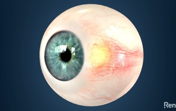
Pterygium
A pterygium (pronounced tur-IJ-ee-um), also known as surfer's eye is a chronic inflammatory response to UV exposure. This fleshy growth extends from the white of the eye onto the clear cornea and can remain small, or grow large enough to interfere with vision. Many patients seek treatment for cosmetic reasons - they simply do not like the look of a pterygium. For others, a pterygium can cause irritation and reduce vision either from traction distorting the cornea, or by encroaching onto the visual axis of the eye itself. A pterygium can often develop from a pinguecula. Infrequently, it represents a form of cancer that requires removal.